


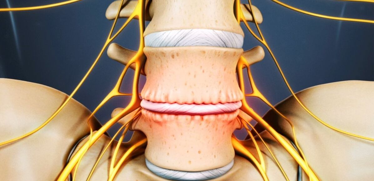
Osteochondrosis is a chronic degenerative-dystrophic disease that develops under the influence of many quite different factors. Initially, pathological changes occur in the nucleus pulposus (the inner contents of the intervertebral disc), and subsequently spread to the fibrous ring (outer shell of the disc) and other elements of the spinal motor segment (SDS). This may be a result of the body’s natural aging process, or it may occur against a background of injuries, increased load on the spine, and other causes. In any case, osteochondrosis is only the first stage of intervertebral disc destruction, and if it is not treated, protrusions and hernias form, which often require surgical removal.
The intervertebral disc is a cartilage formation that separates the vertebral body and acts as a shock absorber.
Lumbar osteochondrosis: what is it
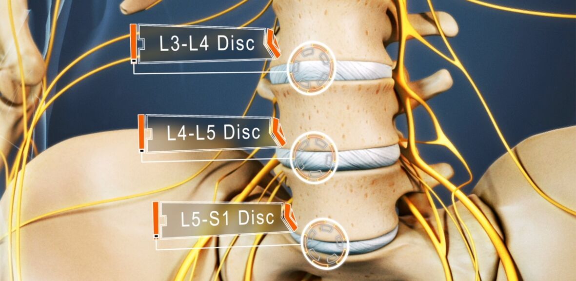
From osteochondrosis suffers from 48 to 52% of people. And osteochondrosis of the lumbar spine is the most common. The disease can affect any intervertebral disc of the lumbosacral spine, some of them, or even all. Often, L5-S1, L4-L5 discs suffer, less often L3-L4. The upper lumbar disc (L3-L2 and L2-L1) is less frequently affected.
The prevalence of lumbar osteochondrosis is due to the fact that the greatest load in the performance of any physical work, especially lifting and carrying weights, walking, running, sitting falls on the lower back. The lumbar spine is made up of 5 vertebrae, which are much larger than the thoracic and cervical vertebrae. Therefore, the intervertebral disc that separates it is larger in size. Usually, the lumbar region has a slight anterior curvature (physiological lordosis). It is the last mobile part of the spine and adjacent to the fixed sacrum, so most often they talk about lumbosacral osteochondrosis.
If osteochondrosis was previously considered an age-related disease, today its first manifestations can already be observed at the age of 15-19 years. Among children in their thirties, already 1. 1% of people experience severe symptoms of degenerative-dystrophic changes in the intervertebral disc. And in representatives of the older age group (from 59 years), the clinical manifestations of the disease are already present in 82. 5%. At the same time, the incidence of pathology continues to grow steadily, which is largely due not only to the increase in the average age of the population of the country, but also to lifestyle changes that are not getting better.
Reasons for development
Today, there is still no consensus on the etiology of degenerative diseases of the spine. However, the main theory of their development is involuntary. According to him, osteochondrosis is the result of previous damage to the intervertebral disc and spinal bone structure, as well as the occurrence of inflammatory and other processes. This theory suggests that degenerative changes are genetically determined and, in fact, unavoidable. And their clinical manifestations, especially in young and middle -aged people, are due to the influence of various endogenous and exogenous factors.
Thus, the development of lumbar spine osteochondrosis is facilitated by:
- heavy physical labor, especially in connection with heavy lifting;
- sedentary, sedentary lifestyle;
- any back injuries, including bruises;
- overweight;
- metabolic disorders;
- violation of posture, deformation of the spinal column;
- flat feet and other foot pathologies;
- pregnancy, especially multiple pregnancies.

Pathogenesis
Regardless of its cause, intervertebral disc degeneration occurs when the intensity of the catabolic process (cleavage and molecular oxidation) of a matrix protein begins to exceed the rate of its formation. One of the main things in this process is the malnutrition of the intervertebral disc.
Since they, like most cartilage in adults, have no direct blood supply, as they have no blood vessels, the supply of nutrients to them and the removal of metabolic products occurs through diffusion with sequential compression and relaxation of the disc during movement. The main structure that provides power to the disk is the end plate located on its top and bottom surfaces.
By itself, the end plate is a bilayer formed by cartilage cells and bone tissue. Thus, their cartilaginous part is adjacent to the disc, and bone - to the vertebral body. They are distinguished by a fairly good permeability, which ensures the exchange of material between cells, material between disc cells and blood vessels that pass through the vertebral body. Over the years, especially with the negative impact of external and internal factors, the structure of the endplates changes, and their blood supply decreases, leading to a decrease in the intensity of metabolism in the intervertebral disc. As a result, its ability to produce new matrices is reduced, leading to a progressive decrease in its density with age.
At the molecular level, this is accompanied by:
- a decrease in the rate of diffusion of nutrients and metabolic products;
- decreased cell viability;
- accumulation of cell decay products and altered matrix molecules;
- a decrease in the production of proteoglycans (high -molecular -weight compounds responsible for the formation of new cartilage cells and which are the main source of chondroitin sulfate synthesis);
- collagen scaffold damage.
Possible consequences
As a result of the constant changes, the intervertebral disc becomes dehydrated, and the nucleus pulposus loses its ability to distribute the load that falls on it adequately. As a result, the pressure inside the disc becomes uneven, and therefore the fibrous rings in some places experience excessive load and compression. Since this happens with every movement of a person, the annulus is always subjected to mechanical stress. This leads to bad changes in it.
Also, often a decrease in the height and elasticity of the disc leads to compensatory changes in the adjacent vertebral bodies. Bone growths called osteophytes form on its surface. They tend to increase in size over time and also merge with each other, excluding the possibility of movement in the affected PDS.
Since the lack of nutrients causes damage to the collagen skeleton, under the influence of the pressure of the nucleus pulposus at a certain point, the normal structure of the fibers that form the fibrous ring is disrupted. If there is no intervention, this eventually leads to cracks and breakage in it. Gradually, more and more fibrous ring fibers at the site of application of the torn pressure, leading to its protrusion. This is mainly facilitated by the increased load on the spine. And because the lumbar region takes the main load during movement and any physical activity, it most often suffers.
The protrusion of the intervertebral disc without rupture of the end of the fibrous ring and with the size of its base more than the protruding part is called protrusion. With a complete rupture in one place or another, an intervertebral hernia is diagnosed.
With partial destruction of the fibrous ring fibers, the pressure in the disc gradually decreases, leading to a decrease in tension and the fibers themselves. This leads to a violation of its fixation and, consequently, the pathological mobility of the affected spinal movement segments.
The vertebral motor segment (SMS) is the structural and functional unit of the spine formed by the intervertebral disc, adjacent vertebral bodies, facet joints, ligaments and muscles attached to this bone structure.
Normal spinal function is only possible with proper PDS operation.
Symptoms of lumbar spine osteochondrosis
The disease can be asymptomatic for a long time, and then begin to manifest itself as a slight discomfort in the lumbar region, gradually gaining strength. But in some cases, lumbar osteochondrosis begins acutely, immediately giving rise to a strong pain syndrome. In most cases, pathological signs first appear after 35 years.
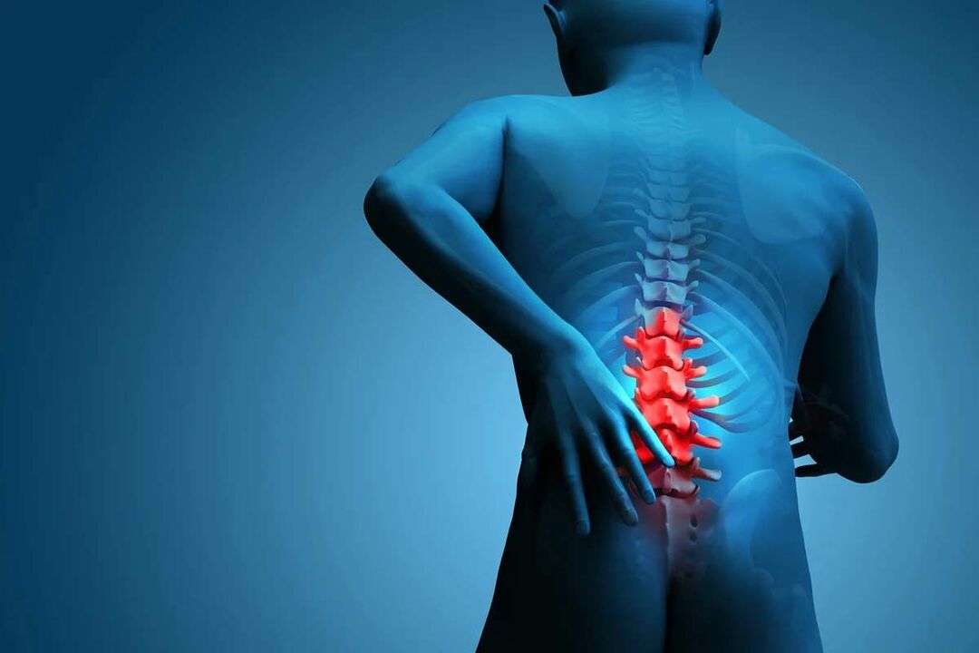
Back pain is the main symptom of this disease. It can vary in character and be both painful and dull, and acute, persistent or episodic. But essentially for pathology, especially in the early stages of development, alternating periods of deterioration and remission are characteristic, and both hypothermia or lifting of heavy objects, or unsuccessful movements, can suddenly trigger another deterioration in well-being.
The pain is often accompanied by numbness and tension in the back muscles. They are exacerbated by physical exertion, sudden movements, weight lifting, bending over, and even coughing and sneezing.
If, due to the instability of the vertebral body, the nerve roots extending from the spinal cord are flanked by one or another anatomical structure, this will lead to the development of appropriate neurological disorders. Their main manifestations are:
- shooting, severe pain radiating to the sacrum, back, lower limbs or perineum;
- sensitivity disorders of varying severity;
- mobility restrictions, lameness;
- weakness in muscles defined by pinched nerves.
In the lumbar spine, the spinal cord ends at levels 1-2 of the vertebrae and enters what is called the cauda equina, formed by the accumulation of spinal roots. Moreover, each of them is responsible not only for the preservation of muscles, but also for certain organs of the small pelvis, so prolonged compression can cause disruption in the work of the corresponding organs. This can lead to the development of impotence, infertility, gynecological diseases, hemorrhoids and other disorders.
The clinical picture of lumbar spine osteochondrosis, especially with its long course and occurrence of spinal root compression, largely depends on the extent of the lesion, i. e. a particular disc that has undergone degenerative-dystrophic changes.
- L3-L4 disc defeat-pain is given to the anterior-inner part of the thigh, lower leg and inner ankle. This is accompanied by a decrease in the sensitivity of the anterior surface of the thigh, a decrease in the severity or loss of the knee jerk, as well as a decrease in quadriceps muscle strength.
- L4 -L5 disc defeat - pain is given from the upper back to the outside of the thighs and lower legs. Less commonly, this is accompanied by the spread of pain to the back of the foot, including 1-3 toes. In this area, there is a decrease in sensitivity and muscle weakness. Occasionally hypotrophy and incomplete extension of the big toe develop.
- Damage to the L5-S1 disc-pain begins in the middle zone of the back and descends to the heel along the posterior or posterior surface of the thigh and lower leg and can capture the outer edge of the foot, such as 4-5 toes. In this area of the lower leg, there is a decrease in sensitivity, and the gastrocnemius and gluteus maximus often decrease in size, which is accompanied by their weakness. If the spinal root pathway at this level of PDS is affected, a decrease or loss of the Achilles and plantar reflexes may be observed.
L1-L2 and L2-L3 discs are rarely affected.
The pain that accompanies the disease blocks a person and significantly reduces his or her quality of life. Since they persist for a long time and recur frequently, if not always present, this inevitably affects the psycho-emotional state. As a result, more than half of the patients show signs of chronic emotional stress, depressive disorders, etc.
Diagnostics
If there are signs of lumbar spine osteochondrosis, you should contact a neurologist or vertebrologist. First of all, the doctor collects anamnesis, which consists of explaining the nature of the complaint, the characteristics of the pain, the conditions for its occurrence and reduction, the characteristics of a person's working life, etc.
The second stage of diagnosis, which is carried out as part of the first consultation with the doctor, is a physical examination. During that time, the doctor assesses the condition of the skin, posture, depth of the physiological curve of the spine, the presence of its curvature, etc. The condition of the muscles surrounding the spine, called paravertebral, must be evaluated, as they are often painful and overly tense, which is the body’s reflex response to inflammation and discogenic pain.
Already based on data obtained during examination and interrogation of patients, neurologists may suspect the presence of lumbar spine osteochondrosis. But to exclude the possibility of concomitant pathology, as well as to confirm the diagnosis and accurately determine the degree of damage, the severity of degenerative-dystrophic changes in the intervertebral disc and the involvement of bone structure, laboratory and instrumental diagnostic methods are required.
Laboratory diagnostics
Analysis of various types does not determine in the diagnosis of lumbar spine osteochondrosis. They are more aimed at assessing the level of inflammatory processes and the detection of concomitant disorders.
Therefore, they can be assigned:
- UAC;
- OAM;
- blood tests for sugar levels;
- blood chemistry.
Instrumental diagnostics
All patients with suspected lumbar spine osteochondrosis were shown to have:
- x -ray of the lumbar spine in two projections - allows you to determine the structure of the bone structure, detect anomalies, osteophytes formed, changes in facet joints, etc. ;
- CT - allows you to detect changes in bone structure in the early stages of development from x -rays, as well as identify indirect signs of osteochondrosis;
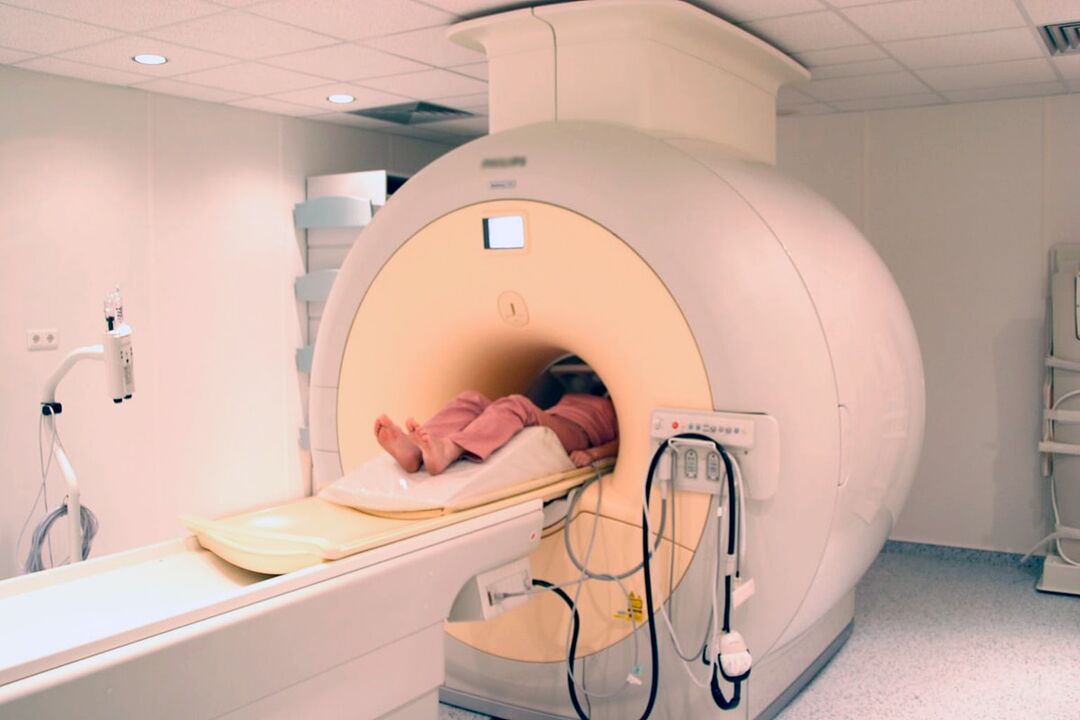
- MRI is the best method for diagnosing pathological changes in cartilage formation and other soft tissue structures, making it possible to detect slight changes in the intervertebral disc, ligaments, blood vessels, and spinal cord and accurately assess its severity and potential risk.
In addition, it may be recommended to:
- densitometry - a method of determining bone density, which makes it possible to diagnose osteoporosis, which is very common in the elderly;
- myelography - allows you to assess the condition of the CSF pathway of the spinal cord and the degree of damage to the protruding disc, which is especially important in the presence of intervertebral hernias that have already formed in the lumbar spine.
Treatment of lumbar osteochondrosis
When diagnosing osteochondrosis, as a rule, initially all patients are prescribed conservative therapy, provided there is no obvious and progressive neurological deficit. But his character is strictly individually chosen.
Because the disease is chronic, and the regenerative capacity of the intervertebral disc is very limited, especially with significant degenerative-dystrophic changes, the main objective of therapy is to stop further progression and eliminate the symptoms that bother the patient. Therefore, treatment is always complex and includes:
- drug therapy;
- manual therapy;
- physiotherapy;
- exercise therapy.
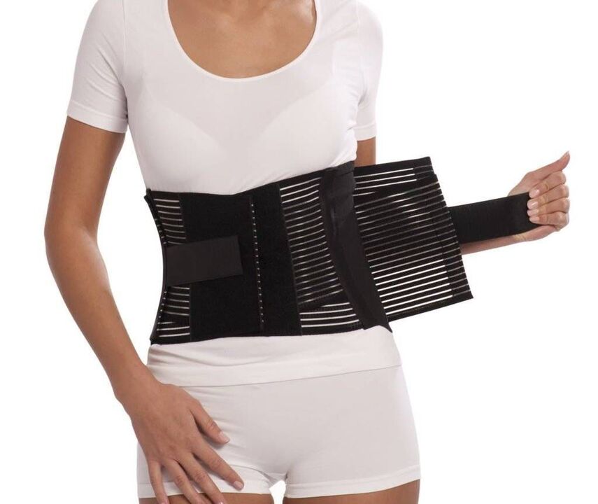
In the acute period, patients are shown to limit physical activity or even adhere to bed rest for 1-2 days. This will help relax the muscles and reduce the pressure inside the discs. If you need to sit, walk or do physical work for long periods of time, you should wear a stabilizing lumbar corset.
After the end of the acute period and during remission of the disease, on the other hand, it is important to move as much as possible, but with caution and exclude increased pressure on the lower back. Patients need to acquire proper sitting skills, lifting objects off the floor, carrying heavy loads, as all this affects the pathological course. It is important to avoid oblique and sudden movements, to lift something off the floor or a low surface, after bending your knees, and not to bend down. You just need to sit with your back straight in a chair that supports your back well. In addition, during sedentary work, it is important to regularly rest for short workouts. It is important to avoid falls, jumps, brisk running and hypothermia.
With osteochondrosis, it is important to maintain weight within optimal limits, and for obesity, diet and physical exercise appropriate to the patient's condition are indicated, as excess weight puts an increased load on the lower back and causes faster development of pathological changes in the disc. .
On average, conservative therapy is usually designed for 1-3 months, although it can last longer. But even after completing the main course prescribed by a doctor, it is necessary to continue taking some medications, exercise therapy and follow the recommendations on lifestyle.
Medical therapy
The main component of drug therapy is individually selected drugs from the NSAID group. When choosing it, doctors take into account not only the severity of the pain syndrome and the course of the inflammatory process, but also the nature of the corresponding disease, especially the digestive tract, because NSAIDs with prolonged use can affect their condition. mucous membranes and trigger exacerbation of various pathologies of the digestive system.
It is necessary to use NSAIDs for acute pain in the lower back, and immediately after its occurrence. Preferably in 1-2 days. Depending on the severity of the patient’s condition, they can be given intramuscularly, in the form of rectal suppositories, local agents and in oral form. The admission period cannot exceed 2 weeks. In the future, individually selected medications are taken on demand, but try to avoid frequent use.
Recently, more often preference is given to drugs, as active ingredients, which include selective inhibitors of cyclooxygenase-2.
Also, patients were given drugs of the following groups:
- muscle relaxant - helps relax overly tense muscles and thus reduces back pain;
- chondroprotectors - improve the course of metabolic processes in the intervertebral disc (especially effective when initiated in the earliest stages of development of lumbar osteochondrosis);
- Vitamin B - contributes to increased nerve conduction;
- antidepressants and anxiolytics - are used for long -term osteochondrosis, which leads to depression, chronic fatigue and other psychological disorders.
With very severe pain, especially of neurological origin, therapeutic restriction is performed. They involve the introduction of an anesthetic in combination with corticosteroids at a point near the compressed nerve, which leads to rapid elimination of pain. But the procedure can only be performed in medical institutions by specially trained health workers, as it is associated with the risk of complications.
Manual therapy
Manual therapy allows not only to improve the quality of blood circulation in the area of influence, but also to significantly reduce the severity and duration of pain in osteochondrosis. It effectively relieves muscle tension and allows you to eliminate functional blocks, which significantly increases mobility in the affected SMS.
Also, through well -carried out manual therapy, it is possible not only to increase the distance between the vertebrae, to return them to the correct anatomical position, but also to release the compressed nerve roots. As a result, the pain is quickly eliminated and the nerve disorder disappears. It also reduces the possibility of complications and disturbances in the work of internal organs.
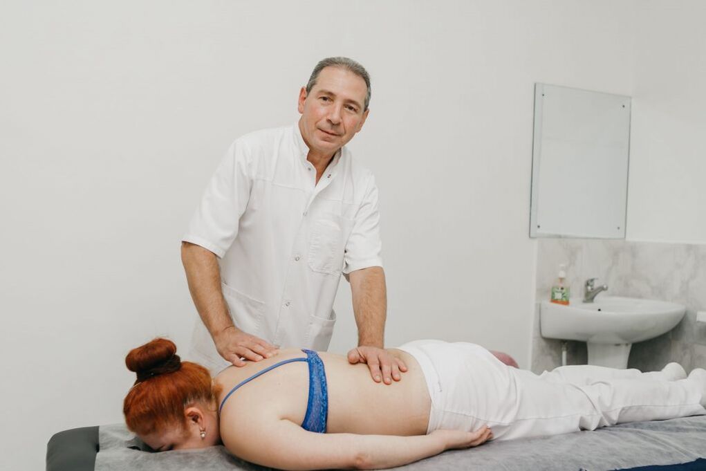
Additional positive features of manual therapy are improving mood, strengthening immunity, activating the body's natural recovery mechanisms and increasing efficiency. Usually after the first session there is a significant improvement in well -being, and in the future the effect becomes more pronounced. As a rule, the course consists of 8-15 sessions, and it is important to complete it to the end, even with complete normalization of well-being.
Physiotherapy
After a decrease in acute inflammation, a course of physiotherapeutic procedures is indicated, which not only helps reduce pain, but also improves microcirculation, nutrition and reparative processes in areas of degenerative-dystrophic changes. Often, patients are prescribed:
- electrophoresis with drug introduction;
- electrical neuromyostimulation;
- ultrasound therapy;
- laser therapy;
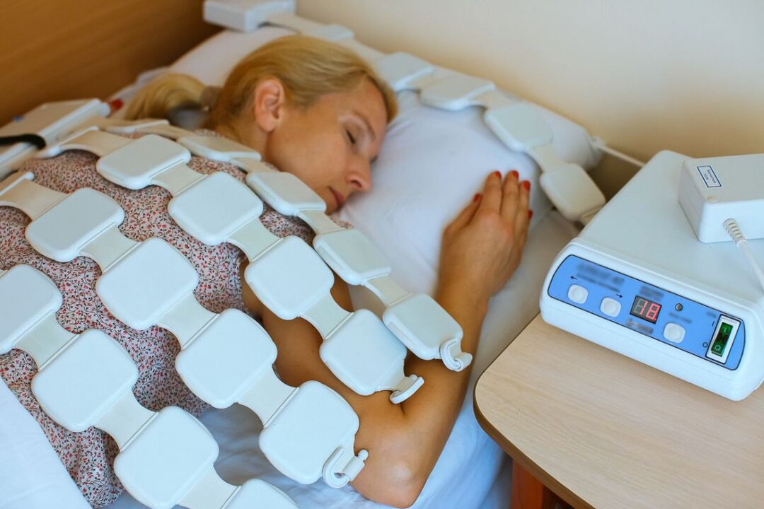
- magnetotherapy;
- UHF.
What specific physiotherapy method will give the best effect, the frequency of its implementation, the duration of the course and the possibility of combining with other types of exposure are determined individually for each patient.
Traction therapy provides excellent results in lumbar spine osteochondrosis. Thanks to him, it is possible to achieve an increase in the distance between the vertebral bodies, which immediately reduces the load on the affected disc. After the session, to consolidate the results, the patient must wear an orthopedic corset.
exercise therapy
After the elimination of acute pain, the treatment program must be supplemented with exercise therapy. Its main task is to stretch the spine and relieve the spasmodic muscles of the lower back. Also, therapeutic exercises help strengthen the muscular corset, create reliable support for the spine and improve posture. In the course of this, blood circulation is inevitably activated and metabolic processes improve, which has a beneficial effect on disc nutrition.
For each patient, a set of exercises was selected individually according to the degree of degenerative-dystrophic changes, the patient’s level of physical development, the nature of the corresponding disorder, age and other factors. Initially, it is recommended to study under the guidance of an experienced exercise therapy instructor.
All patients with degenerative changes in the spine are recommended to visit the pool 2-3 times a week, because swimming lessons minimize the load on the spine, but allow you to strengthen the back muscles effectively.
Therefore, osteochondrosis of the lumbar spine is one of the most common diseases. At the same time, it is able to deprive a person of work capacity for a long time and even lead to disability due to the development of complications. Therefore, it is important not to ignore the first symptoms of the disease, when it is easiest to deal with it. With the appearance of pain, and even more so numbness, limited mobility, back pain, you need to contact a neurologist as soon as possible, undergo the necessary examination and begin treatment. In this case, it is possible to stop the pathological process and return to a normal, full life without significant pain and restriction.